Saurabh Garg1, Somayeh Meysami2,3, Nasrin Akbari1, Rodrigo Solis Pompa1, Thanh Duc Nguyen1, Soojin Lee1, Saqib Basar1, Helen Xu1, Yosef Gavriel Chodakiewitz4, David A. Merrill 2,3,7 Daniel J. Durand4 ,Sam Hashemi1, Cyrus A. Raji5,6
- Vigilance Health Imaging Network, Vancouver, Canada
- Pacific Brain Health Center, Pacific Neuroscience Institute and Foundation, Santa Monica, CA, USA
- Saint John’s Cancer Institute at Providence Saint John’s Health Center, Santa Monica, CA, USA
- Prenuvo Inc
- Washington University School of Medicine in St Louis, Mallinckrodt Institute of Radiology, St. Louis, MO, USA
- Department of Neurology, Washington University in St. Louis, MO, USA
- Department of Psychiatry and Biobehavioral Sciences, David Geffen School of Medicine at University of California Los Angeles, Los Angeles, CA, USA
Abstract
Introduction:
Depression is characterized by persistent feelings of low mood and is often linked to cognitive impairment 1. Neuroimaging studies revealed significant changes in brain volume, particularly in hippocampus and amygdala, areas which play a key role in mood regulation. Exercise has been shown to promote neuroplasticity and improve brain health2, potentially reversing or mitigating the volumetric deficits observed in individuals with depression3. The study examines the relationship between exercise, brain volume changes, and depression, emphasizing the potential of physical activity as a therapeutic intervention to promote mental well-being and improve brain structure in affected individuals.
Methods:
We evaluated 6,458 participants (1,579 depressed, 4,879 non-depressed), across four sites (Vancouver, San Francisco, Los Angeles, Minneapolis) using 1.5T MR imaging with Philips and Siemens scanners. The non-depressed group had a mean age of 52.63 ± 12.62 years (range 21–89), and the depressed group had a mean age of 50.1 ± 12.64 years (range 21–86). We analyzed cortical and subcortical regions, including the hippocampus, amygdala, and cingulate cortex, while correcting for age, sex, and intracranial volume. T1 MPRAGE sequences were processed using the deep learning tool FastSurfer, trained on 134 volumetric T1 images. This method reduced processing time from 8 hours to 35 minutes, enabling large-scale analysis. Physical activity was categorized as moderate to vigorous exercise, defined as activities elevating respiration and pulse rate for at least 10 minutes. Among the depressed group, 1,044 out of 1,593 engaged in moderate or vigorous exercise (439 moderate, 605 vigorous). In the non-depressed group, 1,180 were sedentary, 1,396 moderately active, and 2,302 vigorously active. One-way ANOVA was used to assess brain volume differences across exercise groups after correcting for age and sex, and Spearman correlation identified the top five regions most strongly correlated with exercise. Bonferroni correction was applied for multiple comparisons.
Results:
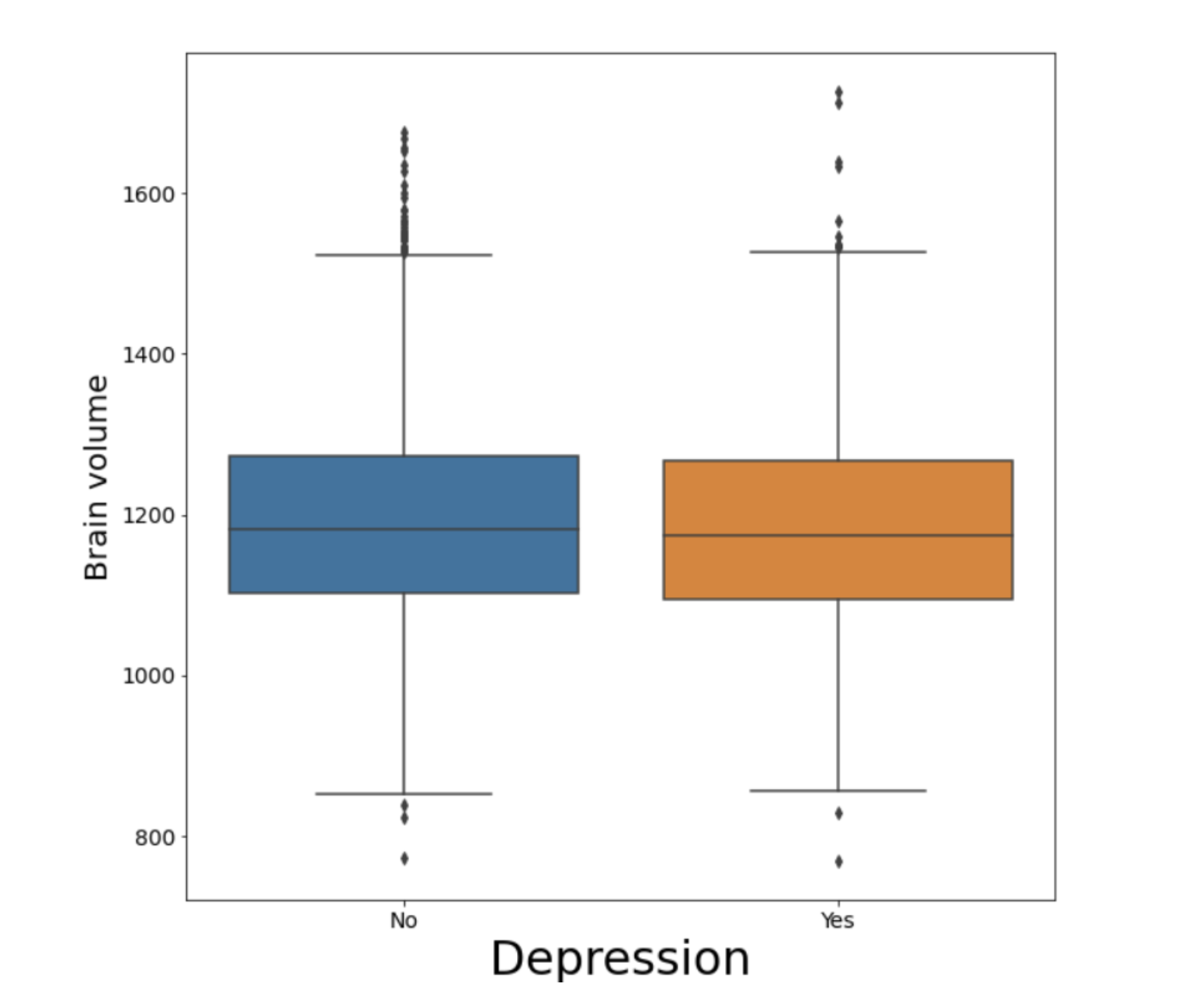
Our results showed significantly lower brain volumes in the depressed population (t = -6.12, p = 9.68e-10) compared to non-depressed controls. Key subcortical regions, such as the hippocampus (t = -6.24, p = 4.57e-10) and amygdala (t = -6.03, p = 1.73e-9), exhibited similar reductions. In cortical regions, significant differences were observed across the cortex, including the cingulate cortex (t = -5.45, p = 5.34e-8), though no significant differences were found in the ventricles, caudal middle frontal cortex, isthmus cingulate, and pars-opercularis.
A one-way ANOVA revealed significant differences in means across the three exercise groups. The results on a few ROIs are shown in Table 1. Further analysis using Spearman correlation revealed the top five brain regions with varying volumes in depressed individuals based on physical activity levels: left rostral middle frontal cortex (r=0.171, p=8.04e-12), left medial orbitofrontal cortex(r=0.165,p=4.26e-11), right middle temporal cortex(r=0.162, p=1.04e-10), and left (r=0.160, p=1.85e-10) and right lingual cortices(r=0.155,p=5.33e-10).
By contrast, the top five regions most strongly correlated with exercise in non-depressed individuals included the left thalamus (0.131, 4.32e-20), right fusiform cortex (r=0.129, p=1.5e-19), right middle temporal cortex (r=0.123, p=6.8e-18), right cerebellum cortex(r=0.119, p=6.46e-17), and left caudal-anterior cingulate cortex (r=0.118, p=1.55e-16).
Discussion:
In our study of 6,458 subjects from the general population, we found that individuals reporting a history of depression exhibited reduced overall brain volumes. Our findings indicate that this reduction is widespread rather than confined to specific brain regions. Several key areas were significantly smaller in the depressed population compared to non-depressed controls.
Additionally, our study explored the impact of exercise on brain volume within the depressed cohort. We found that those who engaged in regular physical activity had larger brain volumes than their sedentary counterparts. This result aligns with previous research suggesting that physical activity is linked to large brain volumes2 and participation in exercise is beneficial for alleviating depression and can lead to a significant reduction in depressive symptoms.
Finally, specific brain areas most strongly linked to physical activity vary between depressed and non-depressed populations. Our results show that the lingual cortex is associated with exercise in depressed individuals, consistent with previous research4. Meanwhile, although less widely studied, the thalamus was also found to be linked to exercise in non-depressed individuals, as reported in5.
Conclusion:
These findings underscore the structural brain changes associated with depression and highlight the importance of exercise in improving the quality of life and reduce brain related changes due to depression.
Acknowledgements:
We appreciate the support and collaboration of the MRI Technologists, Backend, and Patient
Care teams at Prenuvo in the patient care and data acquisition process.
References:
[1] van Tol M, van der Wee NJA, van den Heuvel OA, et al. Regional Brain Volume in Depression and Anxiety Disorders. Arch Gen Psychiatry. 2010;67(10):1002–1011. doi:10.1001/archgenpsychiatry.2010.121
[2] Oyarce DA, Shaw ME, Alateeq K, Cherbuin N. Volumetric brain differences in clinical depression in association with anxiety: a systematic review with meta-analysis. Journal of Psychiatry and Neuroscience. 2020 Nov 1;45(6):406-29.
[3] Raji CA, Meysami S, Hashemi S, Garg S, Akbari N, Ahmed G, Chodakiewitz YG, Nguyen TD, Niotis K, Merrill DA, Attariwala R. Exercise-Related Physical Activity Relates to Brain Volumes in 10,125 Individuals. J Alzheimers Dis. 2024;97(2):829-839. doi: 10.3233/JAD-230740. PMID: 38073389; PMCID: PMC10874612.
[4] Zhao J-L, Jiang W-T, Wang X, Cai Z-D, Liu Z-H, Liu G-R. Exercise, brain plasticity, and depression. CNS Neurosci Ther. 2020; 26: 885–895. https://doi.org/10.1111/cns.13385
[5] Jung J, Kang J, Won E, Nam K, Lee MS, Tae WS, Ham BJ. Impact of lingual gyrus volume on antidepressant response and neurocognitive functions in Major Depressive Disorder: a voxel-based morphometry study. J Affect Disord. 2014 Dec;169:179-87.
[6] Pani, J., Reitlo, L. S., Evensmoen, H. R., Lydersen, S., Wisløff, U., Stensvold, D., & Håberg, A. K. (2021). Effect of 5 Years of Exercise Intervention at Different Intensities on Brain Structure in Older Adults from the General Population: A Generation 100 Substudy. Clinical Interventions in Aging, 16, 1485–1501. https://doi.org/10.2147/CIA.S318679

Fig 1. Boxplot comparing brain volumes between individuals with depression (orange) and those without depression (blue) in a cohort of 6,458 participants (1,579 depressed, 4,879 non-depressed), with non-depressed individuals showing larger brain volumes.

Fig. 2 Boxplot showing significantly larger brain volumes in individuals engaging in vigorous daily exercise compared to those with sedentary or moderate activity levels. The exercise intensity-brain volume pattern is similar across both depressed and non-depressed individuals.

Fig 3. Barplot illustrating the top 5 brain regions correlated with exercise intensity in depressed and non-depressed populations. The lingual cortex is notably correlated with exercise in depressed individuals, while the thalamus shows a stronger correlation in non-depressed individuals.
Table 1. Results of one-way ANOVA comparing brain ROI volumes across three exercise intensity levels, with corrections for age, sex, and intracranial volume (ICV). The analysis shows significant differences (p < 0.05) in all ROIs, indicating that exercise intensity has a significant impact on brain roi volume.

Full Synopsis and Impact:
Motivation: Physical activity may alleviate depression by improving brain health and potentially reversing brain volume deficits.
Goal: To explore the relationship between exercise and brain volumes in individuals with and without depression, focusing on specific regions.
Approach: Analyzed 6,458 participants to assess brain volumes based on physical activity levels. Linear regression and one-way ANOVA examined exercise intensity’s effect on brain volume, controlling for age and sex.
Results: Depressed individuals had smaller brain volumes, while those engaging in moderate to vigorous exercise had larger volumes. Exercise affected different regions, with the lingual cortex in depressed and the thalamus in non-depressed individuals.
Impact: This study highlights the link between physical activity and brain health in individuals with depression, demonstrating that exercise may attenuate volumetric deficits in brain regions, ultimately offering a viable therapeutic strategy to enhance mental well-being and cognitive function.

