Healthcare with humans in mind
We’ve engineered the Prenuvo experience for speed, accuracy, and safety — with your comfort at the core.


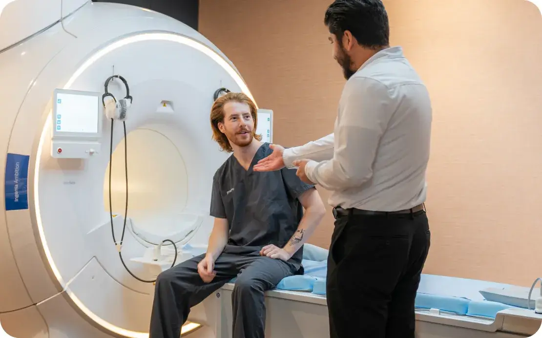
An MRI like no other
Optimized hardware
We work closely with top MRI manufacturers to configure our machines for diagnostic-quality, whole body imaging.
Optimized software
Our software acquisition protocols & calibration provide this multi-parametric diagnostic yield in under one hour.
Future enhancements with AI
We’re actively investigating AI for even better quantification and accuracy, longitudinal tracking over routine scans, and medical research breakthroughs.
We empower our radiologists to give you the very best insights
Every Prenuvo report is carefully interpreted and reported by a radiologist. They’ll also provide helpful context in the report for you and your doctor.
Our radiologists are board-certified physicians with experience in interpreting whole body MRI scans. They review the complete set of images to provide comprehensive reports that consider multiple body systems.
We continually develop our radiology tools specifically for radiologists, making it easier for them to conduct screenings and assess your risks accurately.

Lie back and relax
We’ve designed the scan to significantly reduce anxiety that people might expect from an MRI.
Our machines have a spacious chamber, and you can speak with our technologists at any time during the scan.
You can also listen to auditory beats from HealthTunes or watch your favorite show. Just don’t pick anything with a cliffhanger — you’re all done in under an hour.


The healthcare system needs to catch up
1 in 2 people will develop some form of cancer in their lifetime.1
But only 4 types of cancers are generally recommended for regular screening.2
Yet only 14% of diagnosed cancers are detected by those screenings.3
The other 86% are either not caught by screening or don’t have screening tests.4
Prenuvo scans may help identify certain abnormalities that can be discussed with your healthcare provider, potentially before symptoms appear.
1. Centers for Disease Control and Prevention | 2, 3, 4. NORC at the University of Chicago. (2022). Percent of Cancers Detected by Screening in the U.S. Available at https://cancerdetection.norc.org
Why Prenuvo?
Because we’re voxel visionaries
Voxels are the 3D pixels that our software builds, based on the MRI data we capture. Our expertise on this process is so deep, we’ve literally written the standards on it.
Voxels captured
in 50 minutes
At Prenuvo, we can acquire far more voxels at a diagnostic-quality signal-to-noise ratio.
Different tissue
weights
Prenuvo scans utilize up to 5 times more imaging weightings, providing radiologists with additional data to better differentiate between benign and potentially concerning lesions, thereby helping to minimize false positives.
Number of voxel-averages acquired
Prenuvo scans are designed to provide high-resolution images. Our whole body study includes extensive DWI imaging data, which contributes to the detailed nature of our scans.
New discoveries every day
Follow us on social media to keep up with the latest science and technology of preventative care.


.webp)
*The Prenuvo whole-body MRI screening:
The Prenuvo whole-body MRI screening: (1) can serve as an adjunct to, but is not intended to replace, other established evidence–based screening practices for early detection of specific malignancies (e.g. colonoscopy, dedicated breast imaging, Pap-smear screening for cervical cancer, low-dose chest CT for high risk patients), (2) may not detect some very small cancers that happen to have an MRI appearance very similar to nearby normal tissue structures. We can generally identify cancerous lesions once they are approximately one centimeter in size, although the size limit of detection varies and may be lower in some parts of the body (e.g. the brain) and higher in other parts (e.g. the chest). Our method may not detect lesions in the lining tissue (mucosa) of various body parts, including the mouth, nose, throat, and gastrointestinal tract. As with any medical test, there are limitations, which make it impossible to detect all malignancies and disease conditions, (3)t does not evaluate the heart or heart vessels, (5) does not evaluate detailed lung microarchitecture or pulmonary micronodules, (6) does not replace dedicated breast imaging for screening or diagnostic evaluation (e.g. mammography, breast ultrasound, breast MRI with contrast), (7) is limited in the evaluation of the gastrointestinal tract and does not replace endoscopy or colonoscopy (e.g. cannot detect gastrointestinal polyps), (8) is limited in its assessment of the large joints as the exam is not tailored for detailed evaluation of the joint ligaments, cartilage, menisci, and labrum, (9) should not be considered a primary screening modality of the skin – this is best assessed by direct physical examination, (10) does not visualize smaller brain vessels – as with any imaging test, there are limitations, and stenosis or certain blood flow abnormalities may not be detected, (11) is not intended to replace dedicated diagnostic imaging in the setting of specific clinical diagnostic questions.