Graves' Disease

Axial T2 images of the neck focused on the thyroid, show that it has become diffusely enlarged and swollen, indicative of diffuse thyroiditis. On the current scan (left image), the increased signal on the T2-weighted image highlights the edema, which is sensitive to fluid within the tissue. In contrast, the prior scan showed a smaller and darker thyroid on the T2-weighted image (right image).
Case overview
This case highlights the value of whole body MRI (WB-MRI) in identifying subtle but clinically significant changes in endocrine disorders, helping to improve diagnostic accuracy, and supporting proactive care.
Patient History
A 32-year-old woman with a history of Graves’ disease presented for routine surveillance W-B MRI, with comparison to a baseline scan from two years earlier. Her medical intake form noted nonspecific symptoms, including palpitations, insomnia, anxiety, sensitivity to warm environments, and a mild tremor.
Findings
- Compared to her prior scan, the thyroid gland appeared diffusely enlarged and swollen. This finding, combined with her symptoms, raised suspicion for the interval development of Graves' disease, an autoimmune condition and the leading cause of hyperthyroidism. (Figure 1 & Figure 2)
- No evidence of Graves' eye disease was identified. Complications in the eye are seen in up to 25% of cases.1

Follow-up care
The WB-MRI scan identified thyroid changes and systemic context for her symptoms. We recommended the patient follow up for an endocrinologic evaluation and management.
How the Prenuvo scan impacted patient care:
- Graves’ disease primarily affects women aged 30-60 and if left untreated, can lead to severe complications like thyroid storm (a rare but life-threatening condition in which the thyroid releases too much hormone into the bloodstream) or long-term issues such as Graves’ eye disease.2 Early detection and timely intervention are critical to improving outcomes.
- Our report provided a comprehensive review of other major body systems, which is crucial for identifying complications beyond the thyroid in this multi-system disorder.
- Our WB-MRI is radiation free, which helps avoid additional radiation exposure to the thyroid, a radiosensitive organ, making WB-MRI a safer surveillance option.
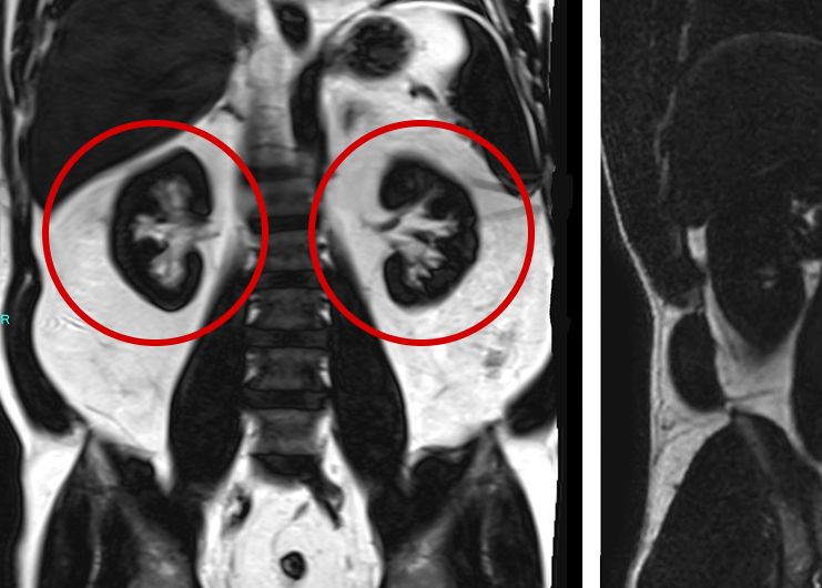
Coronal reformations of whole body diffusion weighted imaging cropped to the center on the head/neck. The thyroid is shown diffusely enlarged and showing increased prominent signal on this series, highlighting the swelling and inflammatory process in this coronal view, compatible with diffuse thyroiditis from Graves’ disease.
References
- Bartalena L, Fatourechi V. Extrathyroidal manifestations of Graves' disease: a 2014 update. J Endocrinol Invest. 2014;37(8):691-700. doi:10.1007/s40618-014-0097-2.
- Antonelli A, Ferrari S, Ragusa F, et al. Grave’s disease: epidemiology, genetic, and environmental risk factors and viruses. Best Pract Res Clin Endocrinol Metab. 2020;34(1):101387. doi:10.1016/j.beem.2020.101387.
Other case studies

Multiple Sclerosis
The Visceral Truth: How Advanced Imaging Detected Severe Internal Fat in an Asymptomatic Man
