Medical imaging plays an important role in modern medicine, allowing the internal structures within the body to be viewed in great detail. Modern imaging techniques such as MRI, X-rays, ultrasound, CT scan and PET scans allow doctors to investigate and diagnose conditions, monitor changes and effects of treatment, and detect any early signs of disease. With more imaging technologies becoming available, healthcare professionals have more options to better assess and evaluate what is happening inside your body.
So, what are the different types of imaging technologies available today?
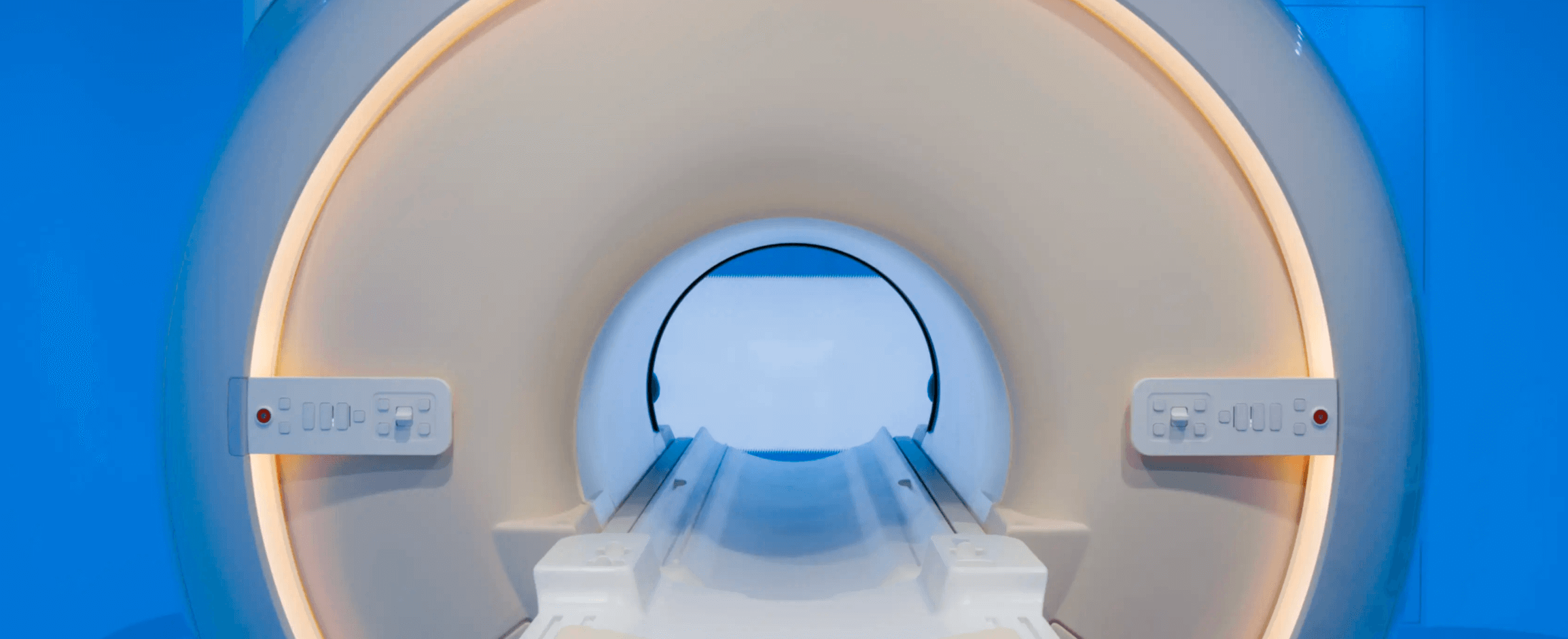
MRI
Magnetic Resonance Imaging (MRI) is an imaging test that uses powerful magnetic fields and radio waves to generate 3D images of the organs, bones and tissues inside your body. Unlike many other imaging techniques, MRIs do not use ionizing radiation. Some MRIs require a contrast medium to provide a clearer image, however Prenuvo’s MRI scans do not use any contrast or dye.
MRIs are best used to image non-bony parts or soft tissues of the body. The brain, spinal cord, organs and nerves, muscles, ligaments and tendons are seen more clearly with MRI than other imaging tools such as X-rays and CT.
Since MRI uses a strong magnetic field, extra caution needs to be taken for people with metallic implants such as pacemakers or deep brain stimulators. However, not all metallic elements in your body prevents you from getting an MRI scan - it is a common misconception. For example, titanium implants and metal tooth fillings are MRI compatible. Ultimately, patients should consult their doctor prior to the examination to determine whether or not they are eligible for a scan.
CT Scan
Computed tomography (CT Scan) is a diagnostic imaging procedure that uses a combination of x-rays and computed technology to produce images of the internal structures of the body. During a CT scan, narrow beams of x-rays are aimed at the patient, producing signals that are processed by the machine’s computer to generate cross-sectional images or “slices”.
Dense structures in the body, such as bones, are easily imaged using x-rays. However, soft tissues can often appear faint. Therefore, when imaging soft tissues with CT, often contrast agents have to be used to ensure the soft tissue is visible in the image. CT scans are used to examine a range of issues including examining blood vessels, diagnosing abdominal issues, investigating tumors, and guiding cancer treatment.
The main risk of getting a CT scan is that it uses ionizing radiation, which has the potential to cause biological effects in living tissue. While getting a CT scan one time may not be harmful, the risk develops with increased exposure to the x-rays over time. However, the risk of developing cancer from x-ray radiation exposure is generally small.
X-ray
An X-ray produces images of the structures inside the body by passing x-ray beams through the body. Different parts of the body absorb varying amounts of x-ray, depending on the density of the material. More dense material such as bone and metal show up as white on x-rays, while fat and muscle appear as shades of gray. Like CT, contrast agents may be used in X-rays to highlight the image of certain bodily structures.
X-rays are a relatively cheap and fast imaging procedure and are best used to examine bones for fractures, infection and tumors. Doctors may also recommend an X-ray to investigate lung infections, heart failure and blocked blood vessels.
Similar to CT, there is a risk associated with ionizing radiation. Generally, the radiation exposure from X-rays are low, and the benefits from these tests far outweigh the risks.
Ultrasound
Ultrasound (also called sonography) is a non-invasive imaging test that uses high-intensity sound waves to produce real-time images and videos of internal organs in the body, particularly soft tissue. The images produced from an ultrasound are called sonograms. Ultrasounds are most commonly associated with pregnancy, however doctors also use ultrasound for diagnosing conditions and guiding medical procedures.
Diagnostic ultrasound can be used to diagnose conditions in the abdomen, kidney, breast and pelvis. Physicians can also use ultrasound to guide needle placement to sample fluid or tissue from tendons, joints, muscles and various organs such as the liver, kidney or prostate.
Unlike X-rays and CT scans, ultrasound uses sound waves to produce images of the anatomy, making it a very safe procedure with no known risks. While there may not be any risks, it still poses some limitations. Since sound waves don’t travel well through air or bone, it’s not the most effective at imaging body parts that have gas in them or are hidden by bone, such as the lungs or brain. For these areas, other imaging tests such as MRI or CT scans would be recommended.
PET Scan
A Positron Emission Tomography (PET) scan is an imaging test that detects metabolic function in your tissues and organs. It requires injecting a safe, radioactive chemical called a radiotracer into a vein within your hand or arm. Diseased cells in your body absorb more of the radiotracer than healthy ones do so the PET scanner can detect this radiation and produce images of the affected tissue.
PET scans are used for a variety of reasons, including assessing the presence of disease and evaluating the function of organs. However, it is mostly used for cancer detection and evaluating cancer treatment. Cancer cells show up as dark spots on PET scans because they have a higher metabolic rate than normal cells. Therefore, PET scans can be useful in detecting cancer, where it has spread, the efficacy of cancer treatment and identifying cancer recurrence.
Like X-rays and CT scans, PET scans also pose a risk due to the use of the radioactive tracer that is injected into the vein. As a PET scan is commonly coupled with a CT scan, the overall dose is relatively high. Therefore, the likelihood of disease has to warrant the scan to be performed and the benefit has to outweigh the risks.
Prenuvo Scan
The Prenuvo scan is a Whole Body MRI scan that is aimed at early detection of disease and cancer. As it is an MRI scan, it does not use any radiation, nor does it use contrast. A Prenuvo scan uses anatomical and functional imaging sequences, and an approach called multiparametric imaging to scan many organs of the body in one session. Due to this technique, Prenuvo scans can capture 10 times more images at high quality compared to traditional MRI.
The primary reason people get a Prenuvo scan is for preventative health measures. When you get a Prenuvo scan, you’re able to obtain a baseline profile of your health that you can use to monitor over time. As we age, our immune system slows down and the risk of disease increases. Those with a family history of disease and cancer, or experience unexplained symptoms over time will use a Prenuvo scan to help identify potential early signs of disease and alleviate uncertainties around what is going on in their bodies.
Like any other MRI scans, the risks mainly surround those with particular metallic implants in the body. While most implants are perfectly safe, patients should consult Prenuvo staff prior to the procedure to determine whether or not they are eligible for a scan.