
Prenuvo Body Composition Analysis


Understand how your body is built and what it’s building toward
Prenuvo’s FDA-cleared Body Composition Analysis helps you better understand how your body is made up by measuring different types of tissue. Using MRI technology, it separates and quantifies visceral fat around your organs, subcutaneous fat under the skin, liver fat, and skeletal muscle. This detailed information can give you a clearer picture of your body’s composition and support more informed conversations about your health and wellness goals.
Your Body Composition Analysis Report is powered by FDA-cleared software that uses AI technology to analyze your results. This report is included with every scan, providing measurements of key body composition details.
Fat analysis
Muscle analysis
Organ volume analysis
What to expect from your Body Composition Analysis Report
Once your scan results are ready, you’ll receive an invitation to review your Prenuvo Body Composition Analysis Report with a Prenuvo provider. During this appointment, you’ll receive clear explanations, annotated images, and easy to understand insights to help you better understand your results. You will receive a copy of your report after completing your Prenuvo Results Review appointment with a Prenuvo provider.
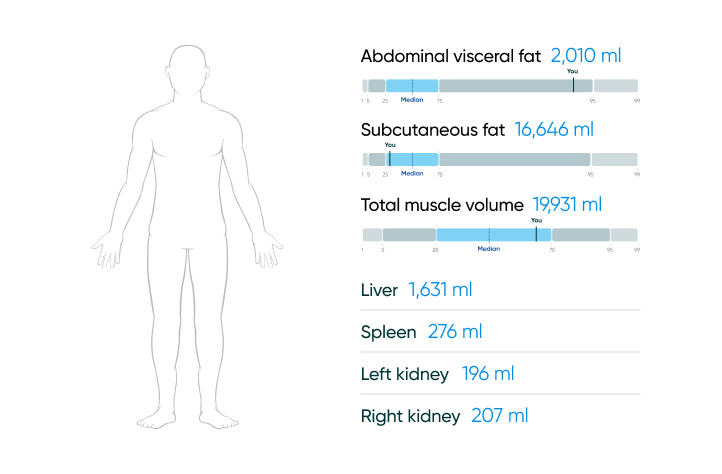
Volume Calculations
Volume calculations for visceral and subcutaneous fat, lower body muscles, and abdominal organs.
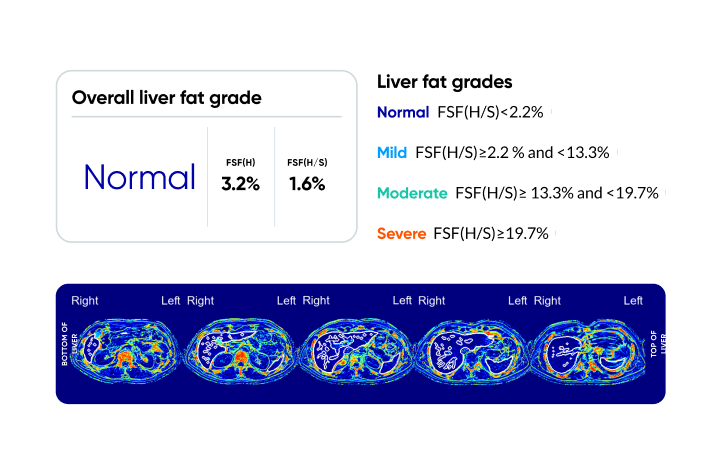
Liver Fat Insights
Liver fat percent and grade, visual heat map of liver fat distribution.
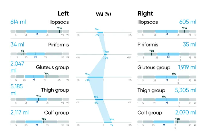
Muscle Asymmetry
Highlights differences between the left and right sides of your lower body, which may influence balance, movement, and overall function.
How to book
Prenuvo Body Composition Analysis
Our proprietary FDA-cleared medical software and AI-learning algorithm that provides a detailed analysis of your body’s muscle mass, symmetry, body fat, and organ volume to help track changes or health insights tied to excess visceral fat or muscle loss.
Includes
- Fat volume analysis (including visceral, subcutaneous and liver fat)
- Muscle volume, with detailed insights into lower body muscle volume and symmetry analysis
- Abdominal organ volume analysis (liver, kidneys, spleen)
Frequently asked questions
The subcutaneous fat calculation includes analysis of the chest / torso and lower body; visceral fat volume analyzes the abdomen.
the liver, stomach, and intestines, considered more dangerous to health than subcutaneous fat (fat just under the skin) as it is pro-inflammatory and can increase the risk of chronic diseases like diabetes and heart disease when present in excessive amounts; it's often referred to as "belly fat" that can't be seen but is located around internal organs. Subcutaneous fat is a type of body fat that is stored just beneath the skin. It is visible and can be felt when pinching the skin, and is distributed through the body. It insulates the body and regulates temperature, provides cushioning and protection for organs and muscles, and stores energy.
Muscle volume / symmetry analysis includes lower body muscles only; volume & symmetry analysis of the chest, core muscles, and arms are not included at this time..
Your percentile values are compared against patients with similar biological sex and age, and normalized by height.
If you have certain medical conditions, the Body Composition report may be inaccurate. These conditions include, but are not limited to, the following:
Hemochromatosis or hemosiderosis of the liver
History of a liver transplant
Situs inversus (a rare condition where major organs are reversed from their normal positions)
Numerous liver cysts, polycystic liver disease, autosomal dominant polycystic kidney disease (ADPKD) with liver cysts, or Caroli disease (a rare disorder affecting the bile ducts)
Sickle Cell Disease or Gaucher’s disease
A muscle-wasting disease or a lipodystrophy
A splenic rupture within the last 12 months"
Our body composition analysis uses MRI to generate body composition measurements, which research suggests1 offers superior accuracy for measurements like visceral, subcutaneous and liver fat volume and muscle volume - without exposure to harmful ionizing radiation. MRI provides a more precise three-dimensional image of soft tissues like fat and muscle, while DEXA primarily measures bone density and provides a less detailed breakdown of body composition. A DEXA scan also exposes patients to a small amount of radiation. While the amount of radiation exposure from a DEXA scan is very low compared to other imaging tests, it can be something to consider if you are getting scanned frequently or seeking to minimize overall radiation exposure.
Source:
1. Comparison between DXA and MRI for the Visceral Fat Assessment in Athletes, Feb, 2022